[ Section Amidella page. ] [ Amanita Studies home. ] [ Keys & Checklist/Picturebooks ]
Amanita clarisquamosa (S. Imai) S. Imai in E.-J. Gilbert"Larger-spored East Asian Amidella"

Technical description (t.b.d.)
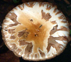
BRIEF DESCRIPTION: The fruiting bodies of A. clarisquamosa are usually medium-sized to large. The cap is 40-100 mm wide, convex to applanate, dirty white to yellowish brown to brownish, with an appendiculate and shortly striate margin; it is covered with brownish to grey-brown, patch-like volval remnants; its context is white, unchanging or barely changing.
The gills of this species are free to subfree, crowded, white to cream-colored but become greyish, grey brown to chocolate brown when dried; and the short gills are truncate and of diverse lengths.
The stipe is 60 - 130 x 10 - 20 mm, subcylindrical to attenuate upwards; its surface is white to dirty white and covered with grey-brown furfuraceous to floccose squamules; the stipe base is not enlarged and basal bulb is not present. The volval remnants form a sac at the base of the stipe; the outer surface of the sac is white to dirty white, and the inner surface is dirty white. The annulus is superior and fugacious.
Spores measure (9.5-) 10.0-13.5 (-14.0) × (5.5-) 6.0-7.0 µm and are ellipsoid to long ellipsoid and amyloid. Clamps are not present on the bases of basidia.
Amanita clarisquamosa was originally described from Japan. It also occurs in China. It grows in mixed forests with broad-leaved trees and conifers.
Amanita clarisquamosa is very similar to A. avellaneosquamosa (S. Imai) S. Imai and A. volvata (Peck) Lloyd. However, A. clarisquamosa differs from A. avellaneosquamosa by its shorter striations on the pileal margin, more densely arranged lamellae and larger spores. The fruit-body of A. volvata becomes reddish brown when cut, and has much thicker subhymenium consisting of 3-4 layers of cells and somewhat smaller spores. Furthermore, the inner layer of the volval remnants consist of a large number of loosely arranged, fusiform, broadly clavate to ellipsoid inflated cells. -- Zhu L. Yang
Photos: Zhu L. Yang (Yunnan, China).
[ Section Amidella page. ] [ Amanita Studies home. ] [ Keys & Checklist/Picturebooks ]
Last change 30 September 2009.
This page is maintained by R. E. Tulloss
Copyright 2005, 2009 by Dr. Zhu L. Yang..
Photographs copyright 2005 by Dr. Zhu L. Yang